
|
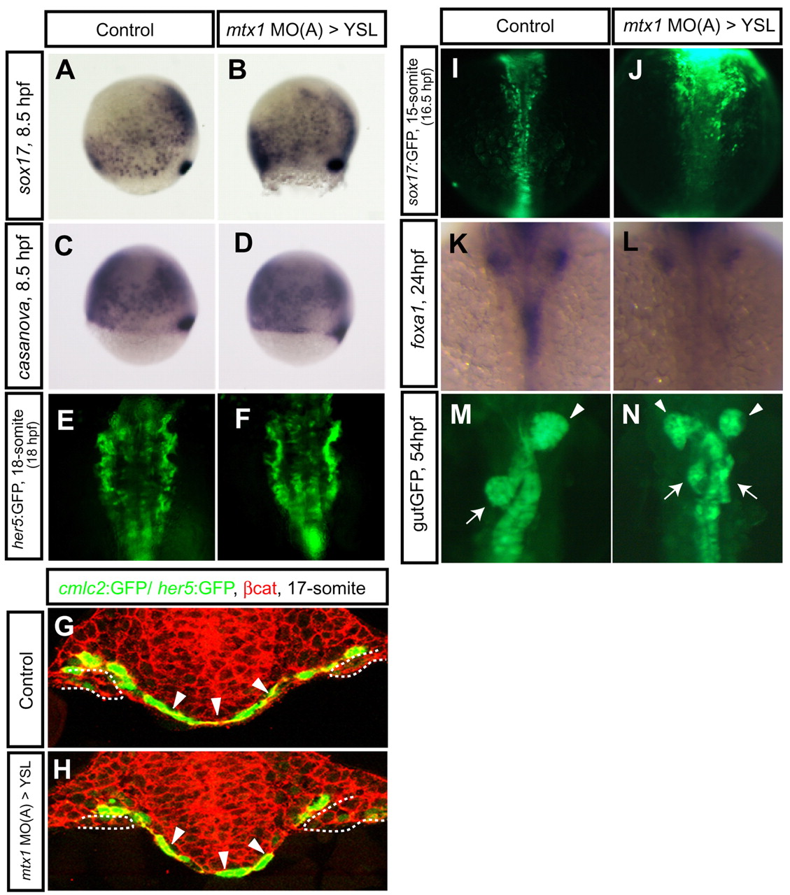
Fig. 3 Endoderm development in mtx1 MO-injected embryos. Endoderm differentiation and morphology in wild-type embryos (A,C,E,G,I,K,M) and embryos injected with mtx1 MO(A) into the YSL (B,D,F,H,J,L,N). Expression of early endoderm markers, sox17 (A,B) and casanova/sox32 (C,D), at 80% epiboly (8.5 hpf) did not appear to be affected in mtx1 MO(A)-injected embryos. (E,F) Anterior views of her5:GFP transgenic embryos at the 18-somite stage show unaffected pharyngeal endoderm development. (G,H) The pharyngeal endoderm was also examined in transverse confocal images (dorsal at the top) of Tg(cmlc2:GFP); Tg(her5:GFP)ne2067 embryos counterstained for β-catenin (red). Arrowheads point to the pharyngeal endoderm and dashed lines outline the myocardial cells in the lateral plate mesoderm. (I,J) Dorsal views of the mid-trunk region of sox17:GFP transgenic embryos at the 15-somite stage. Anterior to the top. sox17:GFP-positive endodermal cells in the future foregut region are coalescing toward the midline in control embryos (I), whereas they appeared to be delayed in their migration in mtx1 MO(A)-injected embryos (J). (K,L) Dorsal views of foxa1 expression in the digestive organ-forming region at 24 hpf. By 24 hpf, endodermal cells have already coalesced at the midline and formed a rod in wild-type embryos (K), whereas they remained as a sheet in mtx1 MO(A)-injected embryos (L). (M,N) Ventral views of gutGFP expression at 54 hpf. Approximately 30% of mtx1 MO-injected embryos showed duplicated hepatic (white arrowheads) and pancreatic (white arrows) buds at this stage (N), further illustrating the endoderm morphogenesis defects.