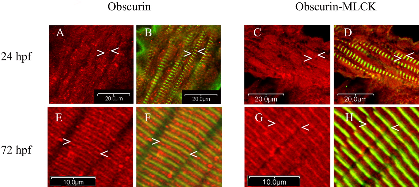
Fig. 3 Cellular distribution of obscurin and obscurin-MLCK in zebrafish skeletal muscle during development. Embryos were fixed at 24 (A-D), and 72 (E-H) hpf and co-immunolabeled with antibodies to the ankyrin binding domain of obscurin (Ank) (red: A,B,E,F) or the carboxy terminal kinase domain of obscurin-MLCK (link7) (red: C,D,G,H) and α-actinin (green: B,D,F,H). At 24 hpf, while most of the cellular obscurin and obscurin-MLCK remains diffusely localized, some begins to organize around the Z (A-D:<) and M bands (A-D:>) of the maturing myofibrils. It is important to note that all myofibrils with a striated pattern of α-actinin staining also demonstrate organization of some of the obscurin and obscurin-MLCK around the M and Z bands. Later in development, by 72 hpf, both obscurin and obscurin-MLCK demonstrate a more distinct striated pattern with obscurin more concentrated at the M bands (E-H:>) and obscurin-MLCK at the Z bands (E-H:<). Similar results were obtained using antibodies to the amino terminal immunoglobulin domains of obscurin (4A8) and the internal kinase domain of obscurin-MLCK (SKII). Scale bars = 20 (A-D) and 10 (E-H) μm.
Image
Figure Caption
Figure Data
Acknowledgments
This image is the copyrighted work of the attributed author or publisher, and
ZFIN has permission only to display this image to its users.
Additional permissions should be obtained from the applicable author or publisher of the image.
Full text @ Dev. Dyn.

