
|
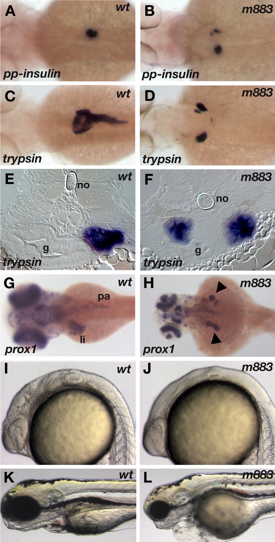
Fig. 1 The m883 mutation causes left-right isomerism of endodermal organs. Formation of endodermal organs in wild-type (A,C,E,G) and m883 mutant embryos (B,D,F,H). A,B: The mutation m883 was initially isolated based on the split appearance of the bilateral preproinsulin expression domain, here in a 3-dpf embryo. C,E: In wild type embryos, as judged from trypsin expression, the right-sided exocrine pancreas extends over several somites along the anterior-posterior axis. D,F: In mutant embryos, the exocrine pancreas is frequently found bilateral and appears shortened. G,H: Expression analysis of prox1 reveals the liver primordium, and demonstrates that liver tissue is also forming on both sides of m883 mutant embryos. I-L: Morphological phenotypes of m883 mutant embryos. Homozygous mutant embryos (J) can be clearly distinguished from wild-type (I) from the 22-somite stage on; the inflation of the brain ventricles fails to occur, while eyes and ears, notochord, and somites appear to develop largely normally. At day 4 (K, L), mutant embryos have severe heart defects, with no blood circulation, causing blood accumulation in the body and retarded growth. Dorsal views in A-D, G, H, with anterior to the left. Transversal sections in E, F, oriented dorsal at top. K,L: Lateral views, anterior to the left and dorsal up. g, gut; li, liver; no, notochord; pa, pancreas.