
|
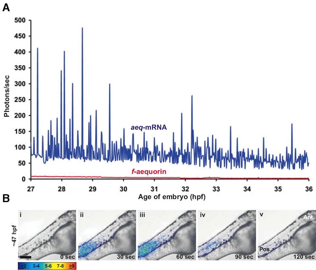
Fig. 8 The use of aeq-mRNA extends the aequorin-based Ca2+ imaging window beyond 24 hpf. (A) A comparison of the aequorin-generated luminescence observed in representative aeqmRNA- and f-aequorin-injected embryos from 27-36 hpf. Traces were generated from data points representing 60 sec of luminescence that were acquired every 60 s for an imaging field covering approximately 64,500 pixels. (B) An example of a localized intercellular Ca2+ signal that is generated in the trunk at ~47 hpf. The panels show the representative pattern of luminescence superimposed on corresponding bright-field images. Each panel displays 60 s of accumulated light with a 30 s interval between each image. Ant. and Pos. represent anterior and posterior, respectively. Color scale indicates luminescent flux in photons/pixel. Scale bar, 200 μm.