
|
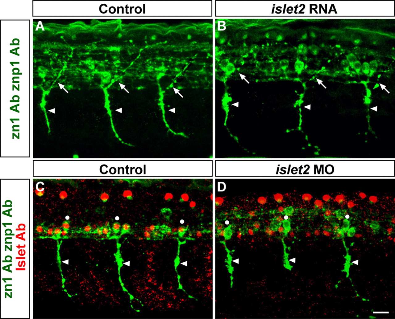
Fig. 6 Formation of CaP subtype identity is independent of Islet2. (A,B) Embryos labeled with zn1 and znp1 Abs (green). Arrows indicate MiP axons, arrowheads indicate CaP axons. At 28 hpf, control embryos (A) have both CaP and MiP axons. Embryos misexpressing islet2 RNA (B) also have normal MiP and CaP axons. (C,D) Embryos labeled with zn1 and znp1 (green), and Islet (red) Abs. Arrowheads indicate CaP axons. CaP cell bodies (dots) co-label with Islet and zn1 Abs and project axons ventrally at 28 hpf in control embryos (C). islet2 MO-injected embryos (D) lack Islet staining in CaP cell bodies and have abnormal CaP axons. Scale bar: 20 µm.