
|
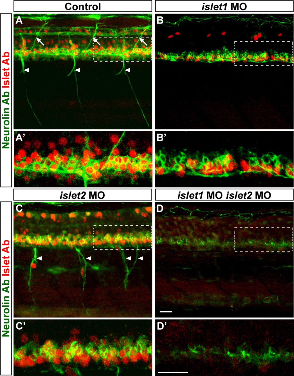
Fig. 3 Islet proteins are required for SMN formation. (A-D) 72 hpf embryos stained with Neurolin (green) and Islet (red) Abs. For each panel, one segment (outlined) is magnified and shown below (A'-D'). Control embryos (A) had dorsally projecting (arrows) and ventrally projecting (arrowheads) Neurolin-positive SMNs and many Islet-positive cells. islet1 MO-injected embryos (B) lacked SMN axons and had 52% fewer Islet-positive cells (*P<0.01, 18 segments of three embryos). islet2 MO-injected embryos (C) had 10% fewer SMN cell bodies (*P<0.01, 24 segments of four embryos). These embryos entirely lacked dorsally projecting SMN axons and ventrally projecting SMN axons (arrowheads) were disorganized. The cells labeled with Islet Ab outside the neural tube next to the ventrally projecting axons are most likely dorsal root ganglion cells that are out of position. Embryos co-injected with islet1 and islet2 MOs (D) lacked all SMNs and 99.5% of Islet-positive cells were absent (*P<0.01, 18 segments of three embryos), although Neurolin-positive floor plate was still present. Scale bars: 20 µm.