
|
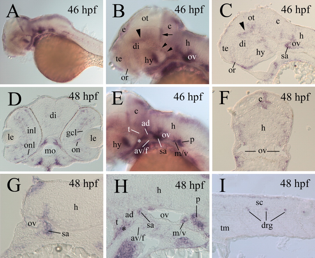
Fig. 5 cadherin-6 expression in 46-48 hpf embryos. A,B,E: Lateral views of whole-mount embryos (anterior to the left and dorsal up), whereas the remaining panels are sections from whole-mount embryos processed for in situ hybridization. C,H,J: Parasagittal sections (anterior to the left and dorsal up). D,F,G: Cross-sections (dorsal up). B,E: Higher magnification images of the anterior half and posterior half, respectively, of the head region of the embryo in A. B: The large arrowhead and two smaller arrowheads point to the cadherin-6 expression domains in the dorsal thalamic and the posterior hypothalamic regions, respectively, whereas the arrow indicates cadherin-6 expression in the anterior cerebellum. E: The asterisk indicates cadherin-6 expression outside the nervous system. C: The arrowhead indicates the dorsal thalamic cadherin-6 expression. G: A higher magnification of the hindbrain at the level of the otic vesicle. H: A higher magnification image of the hindbrain, with the asterisk indicating cadherin-6 expression outside the nervous system. av/f, anteroventral lateral line and facial ganglia; gcl, ganglion cell layer; inl, inner nuclear layer; onl; outer nuclear layer. Other abbreviations are the same as in the previous figures.