
Image
|
Figure Caption
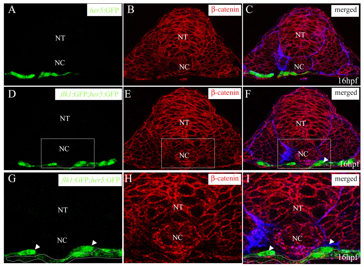
Fig. 2 Angioblasts migrate directly on top of the endodermal layer. Transverse sections of Tg(her5:EGFP)ne2067 embryo (A-C) and Tg(flk1:EGFP)s843; Tg(her5:EGFP)ne2067 embryo (D-I) visualized for GFP (green) (A,D,G), ß-catenin (red) (B,E,H) and fibronectin (blue) (C,F,I). The sections shown are at the level of the 6th somite. White arrowheads mark migrating angioblasts localized right on top of the endodermal layer (outlined by a broken white line). ß-Catenin staining outlines the endodermal layer. NT, neural tube; NC, notochord.
Acknowledgments
This image is the copyrighted work of the attributed author or publisher, and
ZFIN has permission only to display this image to its users.
Additional permissions should be obtained from the applicable author or publisher of the image.
Full text @ Development