
|
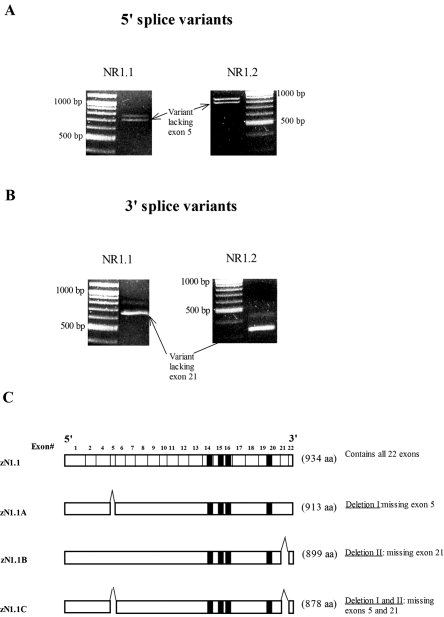
Fig. 5 A: Polymerase chain reaction (PCR) reaction using cDNA from 4-day-old zebrafish. The forward primer lies in exon 4, and the reverse primer in exon 6 of zNMDA R1.1 and zNMDA R1.2 The larger band represents a splice variant, which contains the exon 5 insertion, and the smaller band the splice variant, which lacks this insertion. B: PCR reaction as in A. This strategy represents the C-terminal splice variants produced using a forward primer in exon 20 and a reverse primer in exon 22 of NR1.1 and NR1.2. The larger band represents the splice variant containing exon 21 and the smaller band lacks this exon. C: A diagram representing the three splice variants of zNR1.1, which we detected in zebrafish. ZNR1.2 displays the same splice variants.