
|
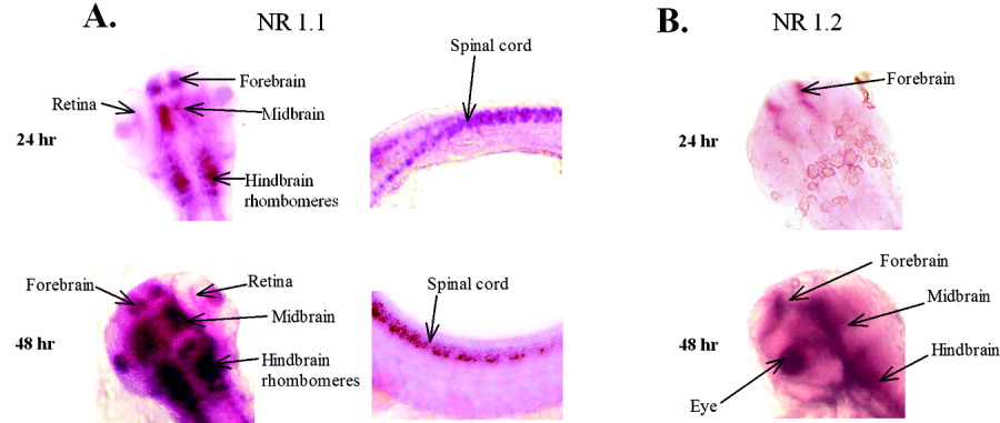
Fig. 6 A: Gene expression of NR1.1 in the head and spinal cord of 24 hours postfertilization (hpf) and 48 hpf. At 24 hpf, NR1.1 is highly expressed in the fore-, mid-, and hindbrain; in the retina; and in the spinal cord. The 24 hpf spinal cord is slightly flexed, presenting a more dorsal view at the rostral end (left), showing bilateral cell labeling. More caudally, the specimen shows that the positive neurons are found both ventrally (top) and dorsally (bottom) in the cord. At 48 hpf, this expression pattern increases in intensity in these same tissues. B: Gene expression of NR1.2 paralog. At 24 hpf, there is light staining in the forebrain. By 48 hpf, all three regions of the brain express NR1.2. The 48 hpf image is a dorsolateral view of the left side of the head. This paralog is not expressed in the spinal cord.