
|
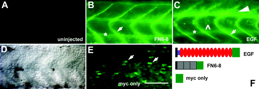
Fig. 4 Injections of epidermal growth factor (EGF) repeats RNA and fibronectin (FN) 6-8 RNA lead to secretion of protein. Whole-mounted 24 hours postfertilization (hpf) embryos were labeled with an anti-myc antibody (orientation as in Figure 1A). A,D: Uninjected embryos are unlabeled (A, corresponding differential interference contrast image in D). E: Overexpression of only the myc tag leads to cellular labeling (arrows). B,C: Overexpression of the FN6-8 RNA (B) and the EGF repeats RNA (C) leads to diffuse labeling of extracellular appearance, concentrated in vertical myosepta (arrows in B,C) and around the notochord (asterisks in B,C). C: After injection of EGF repeats RNA, myc immunoreactivity is additionally concentrated in the ventral spinal cord (arrowhead) and in the region of the horizontal myoseptum (open arrowhead). F: Schematic illustrations of RNA constructs: black rectangles, signal peptide; blue rectangle, cysteine-rich region; red ovals, EGF repeats; gray squares, FNIII domains; green squares, myc tag. Scale bar = 50 μm in E (applies to A-E).