
|
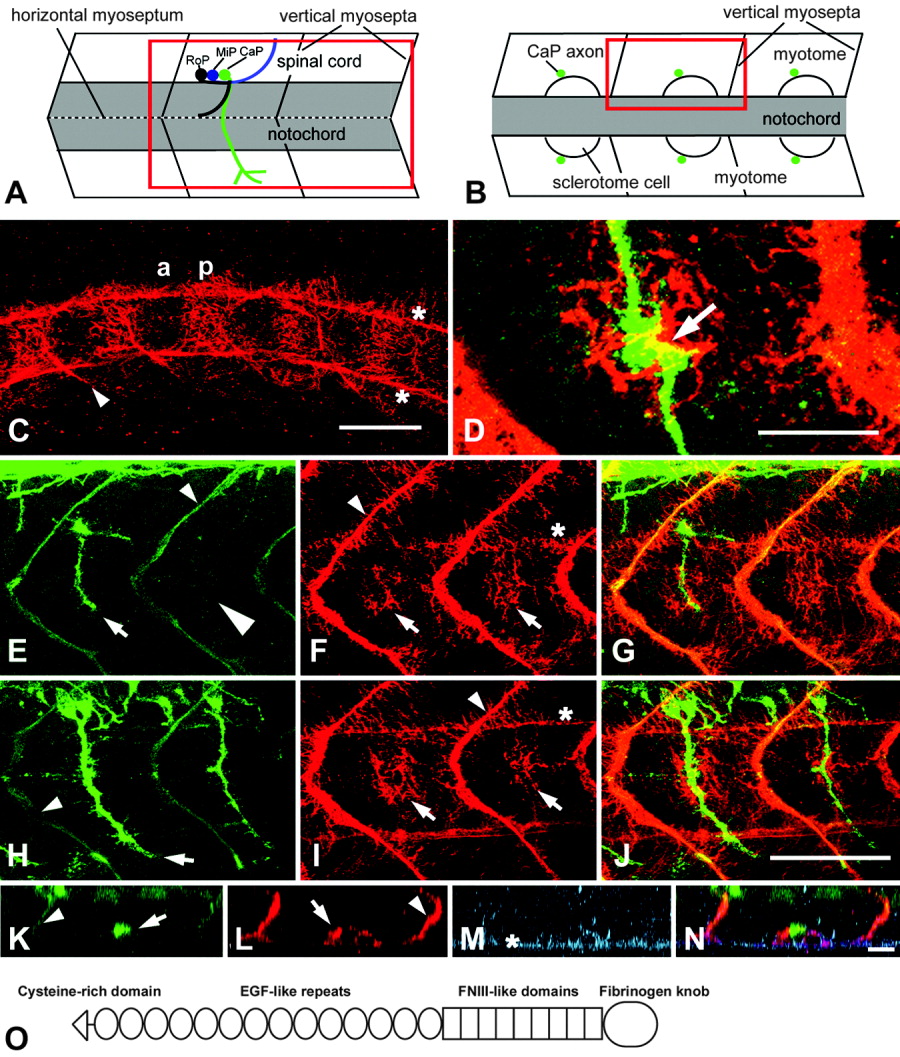
Fig. 2 Tenascin-C immunoreactivity is present in the pathway of motor axons in the region of the horizontal myoseptum. A,B: Schematic representations of trunk segments of 22 hours postfertilization (hpf) embryos from a lateral (A) and a dorsal (B) perspective at the level of the horizontal myoseptum give the orientation of C-J and K-N, respectively; rostral is left. Red rectangles indicate the regions shown. rostral (RoP), medial (MiP), and caudal (CaP) primary motor neurons are indicated for one segment in A. C-J: In lateral views of the trunk at 16 hpf (C), tenascin-C immunoreactivity is found in the posterior (p) but not in the anterior (a) half of the somites in addition to vertical myosepta (arrowhead in C) and the ventral and dorsal edge of the notochord (asterisks in C). D-J: At 22 hpf, double labeling of axons (E,H) and tenascin-C (F,I, overlays in G,J) indicates tenascin-C immunoreactivity in the region of the horizontal myoseptum (arrows in F,I) in the caudal, less-mature (E-G) trunk segments, in which axons have not grown out (large arrowhead in E) or have just reached the horizontal myoseptum (arrow in E), and in rostral (H-J) trunk segments, in which axons have passed the horizontal myoseptum (arrow in H). In D, the overlap of tenascin-C (red) and an axon (green) at the horizontal myoseptum (arrow) is shown at high magnification. K-N: In a horizontally rotated image stack, triple labeled for axons (K), tenascin-C (L), and chondroitin sulfates (M, overlay in N), the ventral motor nerve appears as a dot (arrow in K) next to intense tenascin-C immunoreactivity on the lateral surface of a putative sclerotomal cell (arrow in L). Chondroitin sulfates are labeled on the surface of the notochord (asterisk in M) and more weakly on the surface of the sclerotomal cell. C,E,F,H,I,K,L: Vertical myosepta are indicated by arrowheads (C,E,F,H,I,K,L) and notochord by asterisks (C,F,I,M). O: The domain structure of tenascin-C comprises a cysteine-rich region (triangle), 14 epidermal growth factor-like repeats (EGF-like repeats, ovals), 9 fibronectin type III-like domains (FNIII-like domains, rectangles), and a fibrinogen knob (large oval). Scale bar = 50 μm in C, 25 <μm in D, 50 μm in J (applies to E-J), 15.5 μm in N (applies to K-N).