
Fig. 1
|
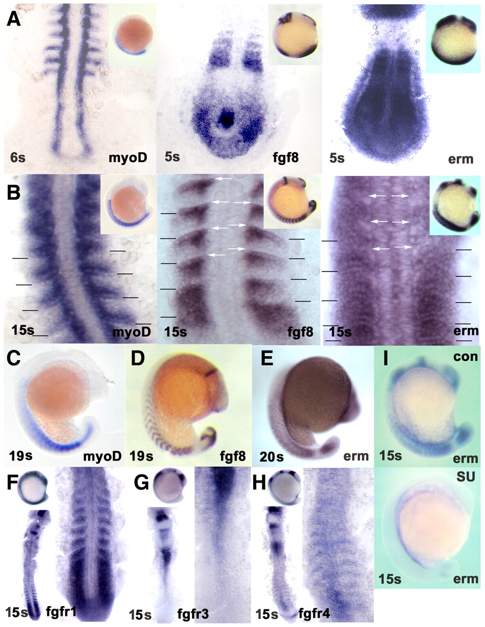
Fig. 1 Fgf8 signalling correlates with myogenic marker expression in the lateral somite. In situ mRNA hybridisation for myod (A-C), fgf8 (A,B,D) and erm (A,B,E,I) at 4-6 s, 15 s and 19-20 s, and fgfr1 (F), fgfr3 (G) and fgfr4 (H) at 15 s. (A,B) Discreet expression of fgf8 overlaps or is immediately adjacent to locations of erm expression in somites. Myod expression parallels fgf8 and erm in the somites, but not elsewhere. Flatmount dorsal view, anterior towards the top. (Insets) Lateral view, anterior towards the top, dorsal towards the left. By 15 s, erm expression is becoming excluded from regions of high fgf8 expression in anterior somites (white arrows). myod and erm expression overlap within the posterior somite. Somite boundaries in the newest five somites are indicated. (C-E) myod, fgf8 and erm expression persist as somites mature. (F) fgfr1 is expressed in presomitic mesoderm and nascent somites. (G) fgfr3 shows little expression in nascent somites. (H) fgfr4 is expressed segmentally in maturing somites and in neural tube. (F-H) Dorsal flatmounts of whole embryo, anterior towards the top, showing tailbuds and newest somites at higher magnification (right). (I) Treatment with SU5402 blocks erm expression throughout the embryo. (C-E,I and insets in F-H) Lateral view, anterior towards the top.