
|
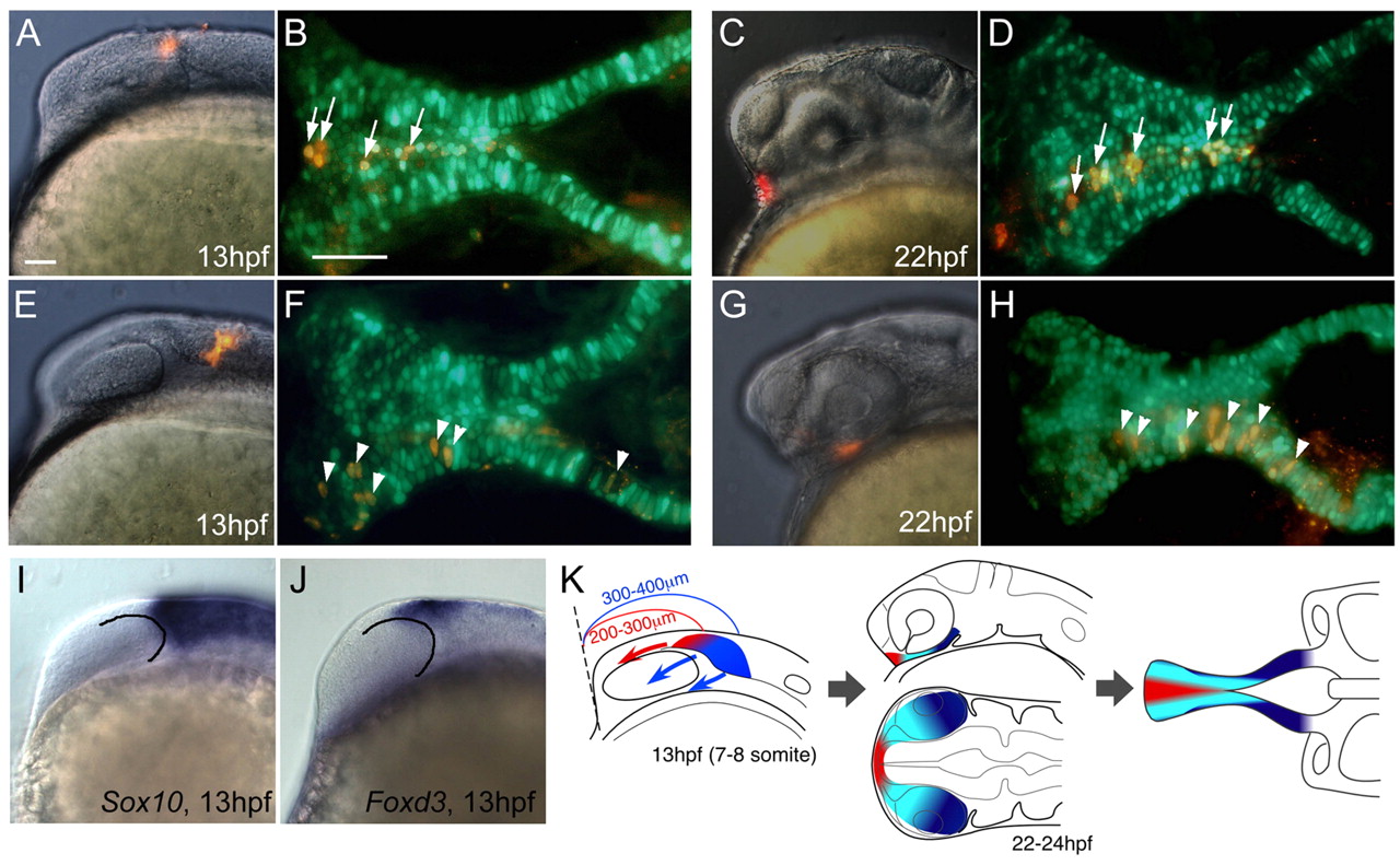
Fig. 4 Fate maps of NC contributions to the ANC. (A,C,E,G) Labeled cells immediately after injection of PKH26, seen in lateral views of living embryos at 13 hpf. (B,D,F,H) Labeled cartilage in the ANC at 80-85 hpf, showing colocalization (yellow) of PKH (red) and sox10:egfp (green), ventral view. (A) Premigratory cranial NC dorsal to the optic vesicle forms median ethmoid (B, arrows). (E) Premigratory NC posterior to the optic vesicle forms trabeculae (F, arrowheads). (C) Postmigratory NC cells anterior to the optic stalk at 22-24 hpf form median ethmoid (D, arrows). (G) Postmigratory NC posterior to the optic stalk form trabeculae (H, arrowheads). (I,J) Expression of sox10 (I) and foxd3 (J) mRNA at 13 hpf, lateral view. Lines delineate the posterior edge of the optic vesicle. (K) Schematic representation of the fate map. In premigratory NC, ethmoid precursors (red) migrate anteriorly, trabecular precursors (blue) migrate laterally and ventrally. Scale bars: 50 μm; in A for A,C,E,G,I,J; in B for B,D,F,H.