
|
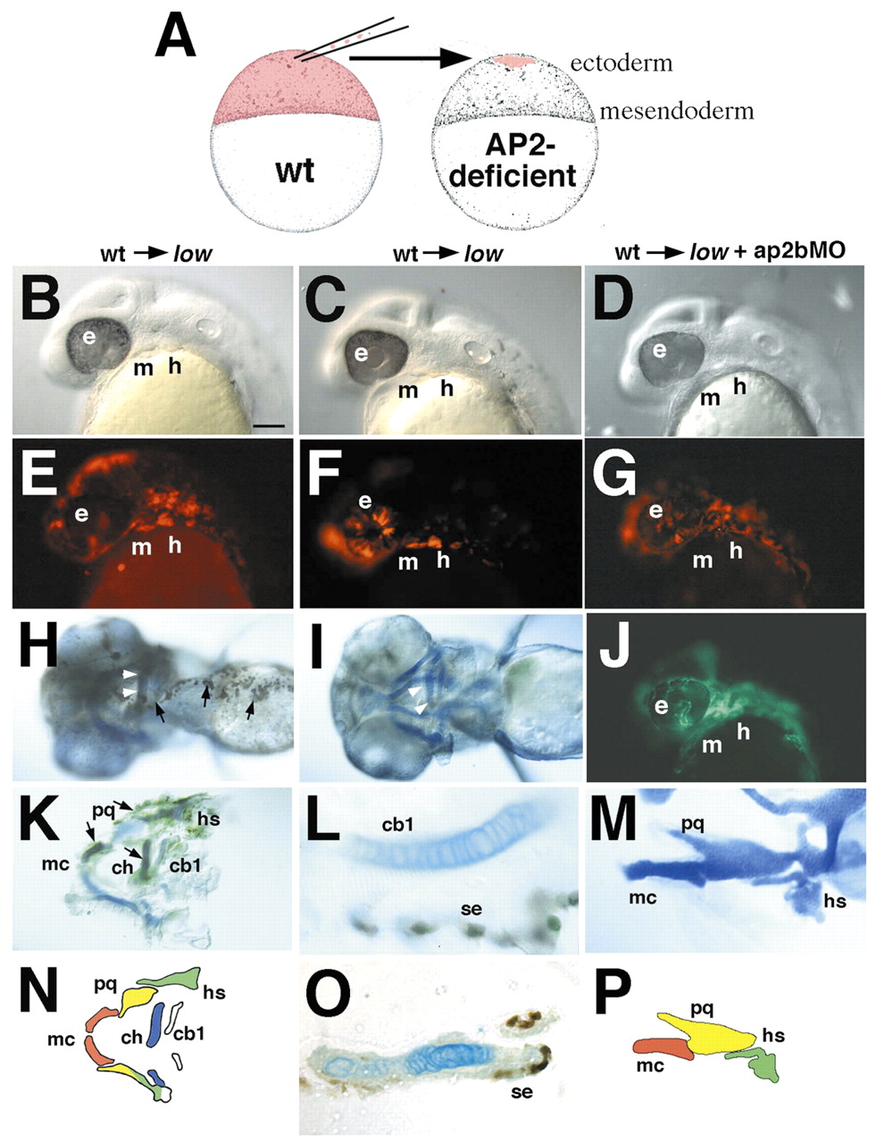
Fig. 8 Transplants suggest that tfap2a and tfap2b are required in the pharyngeal ectoderm, and act non-autonomously in NCCs. Each column represents photos taken at different stages of the same animal. Columns 1 and 2 show transplants from wild-type donors into low mutant hosts, whereas the host in column 3 is a low mutant injected with ap2bMO. Prospective cranial ectodermal cells were transplanted from donor embryos labeled with a lineage tracer into unlabeled hosts at 4-5 hpf (A). Lateral views of living mosaic embryos at 28 hpf (A-G) showing that transplanted cells (red rhodamine-labeled cells in E-G) form patches of ectoderm covering parts of the face and arches. (H,I) Ventral views of mosaic larvae at 4 dpf stained for biotinylated lineage tracer (brown) in the ectoderm. Arrows in H indicate transplanted donor cells in ectoderm. Arrowheads in H,I indicate rescued cartilage of the ceratohyal. (J) fli1-GFP expression in the same embryo as G. (K,M) Flat-mounted rescued cartilages (blue) with ectoderm attached (brown). (L) Higher magnification view of the larvae shown in I, in which transplanted ectodermal cells and cartilage can be clearly distinguished. (O) Section through rescued ceratohyal cartilage in K. (N,P) Diagrams of cartilages shown in K and M. cb, ceratobranchial; ch, ceratohyal; e, ethmoid; h, hyoid arch; hs, hyosymplectic; m, mandibular arch; mc, Meckel's cartilage; ot, otic vesicle; pc, parachordals; pq, palatoquadrate; se, surface ectoderm; t, trabeculae. Scale bars: 50 µm.