
Image
|
Figure Caption
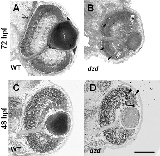
Fig. 3 A-D: Transverse sections on wild-type (A,C) and dazed (B,D) embryos was done at 72 hours postfertilization (hpf) and 48 hpf. Darkly staining pyknotic cells (arrowheads in B,D) were observed near the outer nuclear layer at 72 hpf and at the border of the marginal zone at 48 hpf in dazed embryos. Acellular holes were observed at both 72 hpf and 48 hpf in dazed embryos (asterisks). Scale bar = 80 μm in D (applies to A-D).
Acknowledgments
This image is the copyrighted work of the attributed author or publisher, and
ZFIN has permission only to display this image to its users.
Additional permissions should be obtained from the applicable author or publisher of the image.
Full text @ Dev. Dyn.