
|
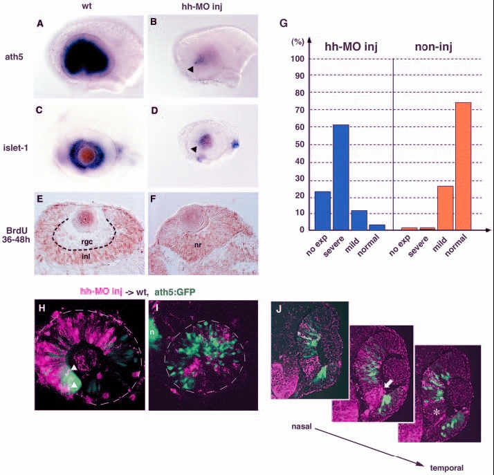
Fig. 7 Shh and Twhh regulate the progression of ath5 expression. (A,B) ath5 expression in 33-hpf wild-type (A) and Hh-MO-injected (B) retinas. ath5 expression fails to progress from the ventronasal retina injected with Hh-MO (arrowhead). (C,D) islet1 expression in 48-hpf wild-type (C) and Hh-MO-injected (D) retinas. islet1 expression fails to progress from the ventronasal retina injected with Hh-MO (arrowhead). (E,F) Labelling of 48-hpf wild-type (E) and Hh-MO injected (F) retinas with an anti-BrdU antibody (brown). Because BrdU is incorporated from 36 to 48 hpf, the RGC layer is BrdU negative in the wild-type retina. By contrast, almost all retinal cells are labelled with the anti-BrdU antibody in the Hh-MO-injected retina. (G) Quantitative assessment of ath5 expression in Hh-MO-injected (blue) and non-injected control (orange) embryos. The defect in ath5 expression in Hh-MO-injected embryos shows a similar profile to that in Gli-MO-injected embryos (blue bars in Fig. 4M). Numbers of Hh-MO-injected and non-injected embryos examined were 33 and 11, respectively. (H) Hh-MO-injected cells were incorporated into 36-hpf wild-type peripheral retina. ath5:GFP expression (green) does not progress towards the Hh-MO-positive area (magenta), although double-positive cells (white arrowheads) are observed at the interface between wild-type and Hh-MO areas. (I) Hh-MO-injected cells were incorporated into 36 hpf wild-type central retina. Hh-MO (magenta) and ath5:GFP (green) are segregated. (J) Three adjacent sections are shown along the axis from the nasal to temporal regions. ath5:GFP expression (green) does not occur in retinal columns derived from Hh-MO-injected donor embryos (magenta). GFP is not expressed even in wild-type retinal columns (white asterisk in right panel) located adjacent to the temporal side of Hh-MO-derived retinal columns (white arrow in middle panel). inl, inner nuclear layer; n, nose; nr, neural retina; rgc, retinal ganglion cell layer.