
Fig. 4
- ID
- ZDB-FIG-260921-25
- Publication
- Reitz et al., 2026 - Molecular characterization of FAM222B as a novel disease gene for dominant cardiovascular laterality defects
- Other Figures
- All Figure Page
- Back to All Figure Page
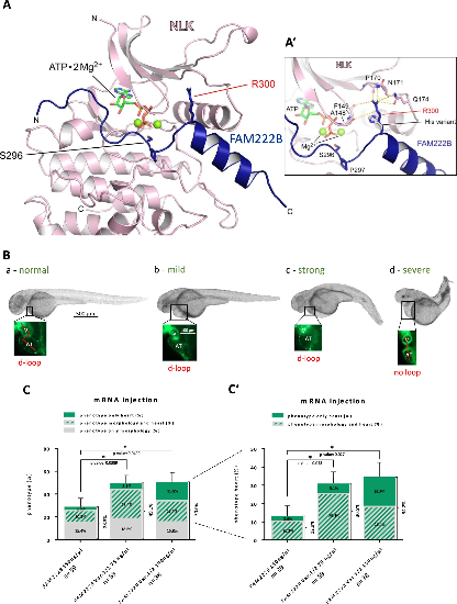
Modelling and functional effects of the FAM222B variant 1/2 rs753661690. (A) Structural model of human FAM222B (residues 286–314) bound to human NLK (residues 140–425). Arg300 (R300) of FAM222B is interacting with the β-sandwich structure of the kinase N-lobe, placing the preceding Ser296 (S296) residue in a position facing the bound nucleotide. Shown is the calculated model where the co-substrate ATP is complexed with two magnesium ions, with the γ-phosphate facing the hydroxyl group of Ser296 of the FAM222B sequence segment S296PISR300. (A’) Close up of catalytic centre showing hydrogen bonds between Arg300 and NLK as well as variant situation showing His300 lacking these. (B) Representative bright field overview and fluorescent heart images of poly(A) mRNA injected wt zfl. After mRNA injection, the 48 hpf old zfl were classified into four different severities of morphological and accompanying cardiac phenotypes: normal, mild (e.g. pericardial edema), strong (e.g. hypotrophic ventricle and concomitant dilated/ hypertrophic atrium) and severe (e.g. no heart looping and no grip). Scale bar: bright field: 500 µm, fluorescent cardiac region: 100 µm. (C) Bar chart classifies zfl (%) into three groups according to the specific developmental disorder: only morphological development (grey) affected, combined cardiac and morphological disorder (grey with green stripes) and only cardiac development (green) affected. N = 3, n = 38–59 in each group, in total. Overall phenotype: two-sided Chi2 test: 150 ng/µl wt against 75 ng/µl variant: p-value: 0.0389, *; 150 ng/µl wt against 150 ng/µl variant: p-value: 0.0499, *. Only morphologically changed zfl: two-sided Chi2 test: 150 ng/µl wt against 75 ng/µl variant: p-value: 0.677, ns; 150 ng/µl wt against 150 ng/µl variant: 0.961, ns. Combined phenotype, including a topical morphological and a cardiac phenotype together in one zfl: two-sided Chi2 test: 150 ng/µl wt against 75 ng/µl variant: p-value: 0.063, ns; 150 ng/µl wt against 150 ng/µl variant: p-value: 0.306, ns. Only cardiac altered zf: two-sided Fisher’s exact test: 150 ng/µl wt against 75 ng/µl variant: p-value: 1, ns; 150 ng/µl wt against 150 ng/µl variant: p-value: 0.056, ns. (C’) Bar chart accumulates all heart phenotypes (combined morphological + cardiac disorder in grey box with green stripes and only cardiac phenotype in green box) and is separately illustrated, meaning taken from (C), to underline differences in the amount of pathologically altered hearts in each group. |