
Fig. 4
- ID
- ZDB-FIG-250714-25
- Publication
- Berger et al., 2024 - Sept10 and sept12 are expressed in specific proliferating cells in zebrafish brain
- Other Figures
- All Figure Page
- Back to All Figure Page
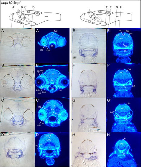
Transverse sections of 4 dpf zebrafish larvae showing detailed sept10 expression in the CNS. A–H: Images of Epon cross sections of 4 dpf zebrafish stained for sept10 transcripts via RNA WISH. Hoechst 33258 staining was used in order to identify single brain regions. Section level and cutting angle are schematically displayed in the drawing above (A′- H′). Note, that the illustration presents the brain scheme of a 5 dpf zebrafish and serve only for rough orientation. Sept10 expression shown in the bright field images was slightly enhanced in ventricular zones (A–C) and particularly prominent in cells at the border of the tectum opticum (TeO) close to the emerging cerebellar plate (CeP in D). Sept10 expression was also detected in cells of the medial medulla oblongate (MO), the otic capsule (OC in F + G) and in ventral mesodermal tissues outside the brain (C–H). Scale bar: 100 μm. See list for abbreviations. Brain schemes were adapted from Mueller and Wullimann, 2016). |
| Gene: | |
|---|---|
| Fish: | |
| Anatomical Terms: | |
| Stage: | Day 4 |
Reprinted from Gene expression patterns : GEP, , Berger, C., Charlotte Kreß, J.K., Helmprobst, F., Sept10 and sept12 are expressed in specific proliferating cells in zebrafish brain, 119387119387, Copyright (2024) with permission from Elsevier. Full text @ Gene Expr. Patterns