
Fig. 4
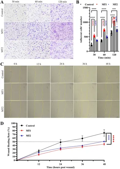
CRIP2 deficiency promotes cell adhesion and impairs cell migration in HUVECs. (A) CRIP2 loss promotes cell adhesion. HUVECs were collected and seeded onto fibronectin-coated plates for 30, 60, or 120 min. The cells were stained with crystal violet. The adherent cells were counted via ImageJ. (B) Quantitative analysis of cell adhesion in CRIP2-MT and control cell lines. Nine microscope fields from each group were randomly captured. The percentages represent the ratios of the number of adherent cells in the CRIP2-MT cell lines to that in the control groups. (C) CRIP2 loss impairs cell migration. HUVECs were collected, seeded and photographed at 0, 12, 24, 36, and 48 h after wounding. Wound closure was investigated via ImageJ. (D) Quantitative analysis of cell migration in CRIP2-MT and control cell lines. Six microscope fields from each group were randomly captured. Two-way ANOVA; #### or ****, P < 0.0001 |