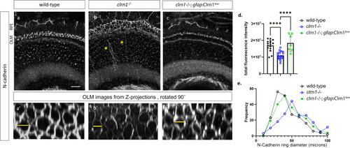
FIGURE

Fig 9

ID
ZDB-FIG-250321-17
Publication
Nonarath et al., 2025 - The USH3A causative gene clarin1 functions in Müller glia to maintain retinal photoreceptors
Other Figures
All Figure Page
Back to All Figure Page
Fig 9

Expression of N-cadherin is reduced in clrn1-/- zebrafish.

Confocal microscope images of N-cadherin immunostaining in transverse cryosections of retinas from 7 dpf (a) wild-type, (b) clrn1-/-, and (c) clrn1-/-Tg(gfap:Clrn1low). Images of N-cadherin staining in the OLM (a’, b’, c’) were generated by rotating projected Z-stacks 90˚at the level of the OLM. The OLM and RPE are denoted to the left of panel (a). Yellow asterisks in (b) indicate delocalized N-cadherin within the inner nuclear layer. (d) Quantitation of N-cadherin fluorescence intensity from 300 um2 central retinal regions revealed that N-cadherin staining was reduced in clrn1-/- but not in clrn1-/-; gfap:Clrn1low (n=12 eyes each genotype). (e) N-Cadherin ring diameter frequency distribution for each genotype. Example measurements marked by yellow lines. Date points in (e) represent 35 rings scored in 300 um2 central retina region (n = 12 eyes each genotype). (****p<0.001; One-way ANOVA). Error bars=SD. Scale Bar=10 mm.

Expression Data

Expression Detail
Antibody Labeling
Phenotype Data

Phenotype Detail
Acknowledgments
This image is the copyrighted work of the attributed author or publisher, and ZFIN has permission only to display this image to its users. Additional permissions should be obtained from the applicable author or publisher of the image. Full text @ PLoS Genet.