
Fig. 2
- ID
- ZDB-FIG-250122-3
- Publication
- Matsumoto et al., 2024 - Foxo3-mediated physiological cell competition ensures robust tissue patterning throughout vertebrate development
- Other Figures
- All Figure Page
- Back to All Figure Page
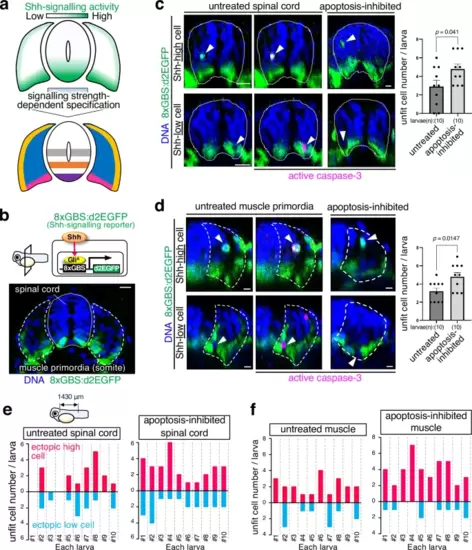
Shh-unfit cells are apoptotically eliminated.a The strength of Shh signalling activity mediates distinct cell types specification in the spinal cord and muscle. b In a horizontal plane, 8xGBS:d2EGFP drives destabilised enhanced green fluorescent protein (d2EGFP) expression in response to Shh signalling activation in the developing spinal cord and muscle of reporter larvae (cross-sectional view with the dorsal on top, 24 hpf). The dotted line and dashed line represent the spinal cord and muscle primordia boundaries, respectively. Scale bar = 10 μm. c, d Caspase-3 activation in cells with impaired Shh signalling activity during spinal cord (c) and muscle development (d). Whole-mount immunostaining of d2EGFP (green) horizonal plane and active caspase-3 (magenta) in Tg(8xGBS:d2EGFP) zebrafish larvae untreated or apoptosis-inhibited (bcl2a mRNA-injected). Arrowheads indicate cells with abnormal Shh signalling-reporter activity. Scale bar = 10 μm. The bar plots show the mean + SEM of unfit cell frequencies in untreated and apoptosis-inhibited larvae. An unpaired two-tailed t-test was used for the statistical analysis. e, f Inhibiting apoptosis enhances the Shh-unfit cell accumulation in the spinal cord (e) and muscle (f). The bar plots show unfit cell frequencies in untreated and apoptosis-inhibited larvae. Each larva has a different number of spontaneously appearing unfit cells with abnormally high or low Shh signalling activity. Source data are provided as a Source Data file. |