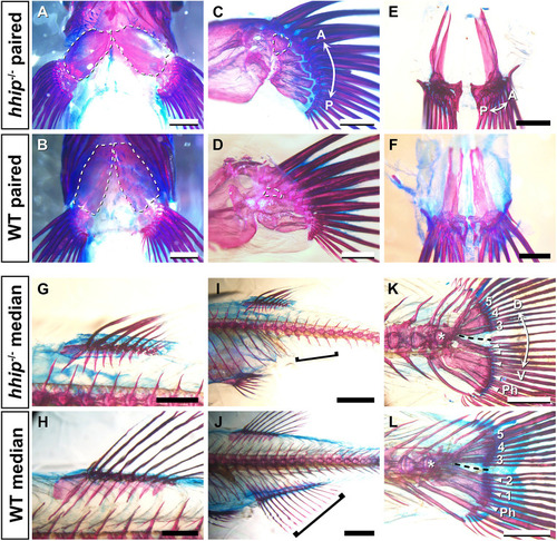
Paired and median fin skeletons in hhip−/− zebrafish. (A,B) Ventral views of the pectoral fin skeleton of hhip−/− (A) and WT (B) zebrafish. White dashed lines indicate the pectoral girdles. (C,D) Medial views of the pectoral fin skeleton of hhip−/− (C) and WT (D) zebrafish. White dashed lines indicate the first proximal radial (PR1). (E,F) Pelvic fin skeletons of hhip−/− (E) and WT (F) zebrafish. (G,H) Dorsal fin skeletons of hhip−/− (G) and WT (H) zebrafish. (I,J) Anal fin skeletons of hhip−/− (I) and WT (J) zebrafish. Black brackets indicate the post-anal region where the anal fin is formed. (K,L) Caudal fin skeletons of hhip−/− (K) and WT (L) zebrafish. Black dashed lines represent the hypural diastema, and the white arrowheads indicate hypurals in the region ventral to the hypural diastema. Numbers indicate the first to fifth hypurals. The white asterisk marks the first ural vertebra. Ph, parhypural. All observations were performed on five hhip−/− and five WT fish. Double arrows indicate the anterior (A)-posterior (P) axis and the dorsal (D)-ventral (V) axis. Scale bars: 500 µm (C-F); 1 mm (A,B,G,H,K,L); 2 mm (I,J).
|
