
Fig. 5
- ID
- ZDB-FIG-240703-107
- Publication
- Liu et al., 2024 - Knockdown of best1 Gene in Zebrafish Caused Abnormal Neuronal and Skeletal Development -A Subtype of Craniovertebral Junction Malformation
- Other Figures
- All Figure Page
- Back to All Figure Page
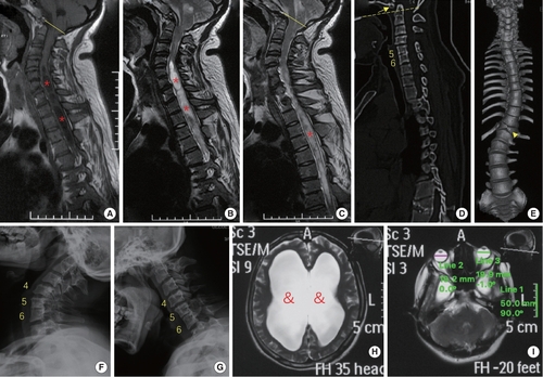
phenotypes of the patient with a premature stop codon of BEST1 gene (p.S79Ffs*153). (A–C) Sagittal magnetic resonance imaging (MRI) revealed Chiari malformation (cerebellar tonsil exceeding the foramen magnum, yellow line) and extensive syringomyelia (red stars) at cervical and thoracic segments. (D–G) Computed tomography and x-ray showed assimilation of atlas (arrow), basilar invagination (odontoid process above the chamberlian line, yellow dotted line), Klippel-Feil malformation (fusion of C5 and C6 vertebrae), butterfly vertebra at T12 (arrow head) and scoliosis. (H, I) MRI of the head indicated hydrocephalus (&) and microphthalmia (diameter of eyeball is smaller than 20 mm). |