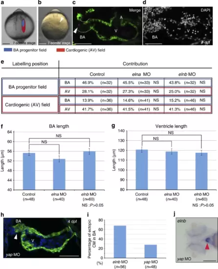
Cell fate determination is altered in elnb morphant BA. (a,b) Fate map of BA (blue) and cardiogenic (red) progenitors in anterior lateral plate mesoderm according to Hami et al.45 Scale bars, 200 μm. (c,d) Example of fate mapping result by injection of fluorescent tracer DiI. Arrowhead indicates injected DiI. Scale bars, 50 μm. (e) Result of cell lineage tracing experiment. In all cases, BA progenitor field to BA or cardiogenic and cardiogenic progenitor field to BA or cardiogenic in both elna and elnb morphants, no statistically significant differences were observd. NS: P>0.05. (f,g) Lengths of BA and ventricle in control, elna and elnb morphants. In all cases no statistically significant differences were observed. NS: P>0.05. (h) Anatomy and histology in yap morphant hearts at 4 dpf. Ecotopic cardiomyocytes are observed in BA (arrowhead). Scale bar, 50 μm. (i) Percentage of ectopic cardiomyocytes in the BA of elnb morphants (control) and yap morphants. (j) elnb expression in yap morphants. elnb expression is not changed in yap morphants (arrowhead). Scale bar, 200 μm. Values are reported as mean±s.d. and P values were determined by the t-test.
|
