
Fig. 5
- ID
- ZDB-FIG-230830-12
- Publication
- Zhong et al., 2022 - Exogenous iron impairs the anti-cancer effect of ascorbic acid both in vitro and in vivo
- Other Figures
- All Figure Page
- Back to All Figure Page
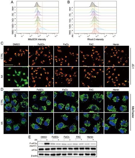
Iron protected AA-induced mitochondrial dysfunction and ER stress. (A-D) A549 cells were treated with AA (2 mM, 2 h) with or without pretreatment of FeSO4, FeCl3, FAC, or hemin, respectively. Then (A) mROS and (B) mitochondria Ca2+ were detected with flow cytometry, (C) MMP was evaluated with JC-1 staining, (Bar = 100 μm), (D) mitochondria swelling was monitored with MitoTracker Green. (Bar = 10 μm). (E) A549 cells were treated with AA (2 mM, 2 h) with or without pretreatment of FeSO4, FeCl3, FAC, or hemin, respectively, and P-eIF2α, eIF2α were detected. β-actin served as the loading control. (For interpretation of the references to colour in this figure legend, the reader is referred to the web version of this article.) |