
Fig. 4
- ID
- ZDB-FIG-230809-10
- Publication
- Prendergast et al., 2023 - CSF-contacting neurons respond to Streptococcus pneumoniae and promote host survival during central nervous system infection
- Other Figures
- All Figure Page
- Back to All Figure Page
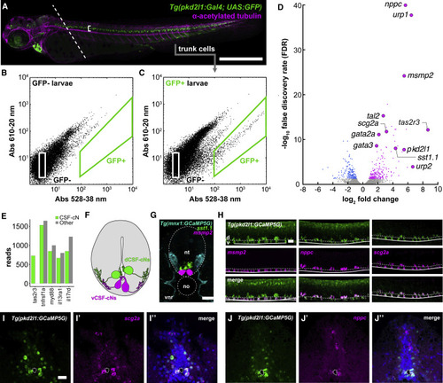
Figure 4. The CSF-cN transcriptome reveals the expression of taste receptors and immune-related secreted factors (A) 3 dpf zebrafish Tg(pkd2l1:GAL4; UAS:GFP) larva immunostained for GFP (green) and acetylated tubulin (magenta). Dashed line indicates plane of decapitation prior to dissociation (anterior tissues were discarded to exclude labeled cells in brain and heart); bracket indicates CSF-cN domain. Scale bars, 500 μm. (B) Calibration FACS plot from non-transgenic siblings. (C) FACS plot from a typical dissociation of cells from transgenic larvae. A small fraction (∼0.2% of input) of cells is green-shifted. Figure S3 quantifies the upper bound of cells/larvae we could reasonably expect to obtain. In Figure S4A, we use qPCR to show that this fraction appeared to be CSF-cNs. (D) Volcano plot of all RNA-seq replicates: 202 transcripts were enriched in CSF-cNs. Figure S4B provides more categorical information on these hits. Figure S6 shows that knocking out individual CSF-cN-enriched taste receptors identified by this approach did not impair CSF-cNs’ ability to detect bitter compounds. (E) Quantification of reads for selected immune-related receptors in both GFP+ CSF-cNs and GFP− (i.e., all other) cells. (F) Schematic representation of a transverse section of the spinal cord and central canal showing the disposition of dorsolateral CSF-cNs (dCSF-cNs, green) and ventromedial CSF-cNs (vCSF-cNs, magenta). (G) A mixed fluorescence in situ hybridization/immunofluorescent stain showing the disposition of motor neurons (cyan, Tg(mnx1:GCaMP5)), dCSF-cNs (green, sst1.1), and vCSF-cNs (magenta, msmp2). The section here is a frontal section, e.g., a cross-section of the spinal cord. Scale bars, 50 μm. (H) Fluorescence in situ hybridization reveals that the putative immune-related secreted factors msmp2, nppc, and scg2a are expressed in CSF-cNs. These confocal stacks are lateral stacks, so at 90° orientation to the image in (G). Scale bars, 50 μm. Figure S5 shows additional in situ hybridization results from some of these RNA-seq hits; most hits we assessed were verified histologically. (I) Transverse section of adult Tg(pkd2l1:GCaMP5G) spinal cord; expression of scg2a is specific to CSF-cNs. Scale bars, 50 μm. (J) Adults continue to express nppc and scg2a in CSF-cNs. See also Figures S3–S6, Table S1, and Data S1. |