
Figure 5
- ID
- ZDB-FIG-230529-49
- Publication
- Cuevas et al., 2023 - Cytoskeletal Keratins Are Overexpressed in a Zebrafish Model of Idiopathic Scoliosis
- Other Figures
- All Figure Page
- Back to All Figure Page
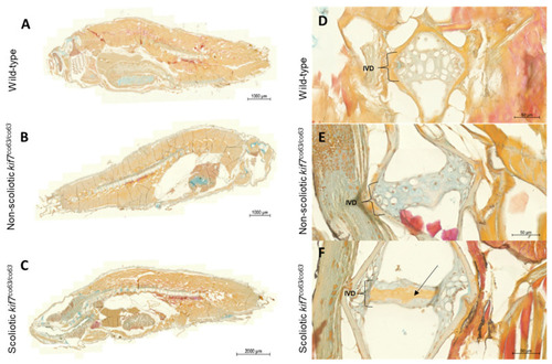
Keratin staining of wild-type (A,D), non-scoliotic (B,E), and scoliotic kif7co63/co63 (C,F) 6 wpf zebrafish (D. rerio). Keratin staining of whole cross section fish (A–C) and selective caudal vertebrae (D–F) depicting the intervertebral disk (IVD) are shown. The IVD is distinguished by vacuolated cells clearly in the wild-type and non-scoliotic kif7co63/co63 fish, but vacuolated cells are less visible in scoliotic kif7co63/co63. Blue: glycosaminoglycans; orange to red: prekeratin to keratin; brown: nuclei. Increased keratin/prekeratin can be seen in the IVD of scoliotic fish compared to non-scoliotic fish and wild type, as denoted by the black arrow. N = 3 zebrafish were analyzed for each group (wild type, scoliotic kif7co63/co63, and non-scoliotic kif7co63/co63). |
| Fish: | |
|---|---|
| Observed In: | |
| Stage: | Days 30-44 |