
Fig. 3
- ID
- ZDB-FIG-230421-22
- Publication
- Lysko et al., 2022 - Unmyelinated sensory neurons use Neuregulin signals to promote myelination of interneurons in the CNS
- Other Figures
- All Figure Page
- Back to All Figure Page
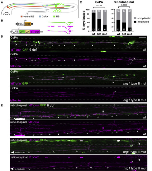
nrg1 type II is required for normal myelination of diverse neuronal classes (A) Illustration of spinal cord neurons assessed with the myelin reporter. RS neuron cell bodies reside in the hindbrain and project posteriorly toward the tail (orange). CoPA neurons reside in the dorsal spinal cord and project anteriorly to the hindbrain (blue). RB sensory neurons reside in the dorsal spinal cord, receive touch stimulation via their sensory arbor, and project both anteriorly to the hindbrain and posteriorly (green). (B) The UAS:GFP-2A-tdTomato-contactin myelin reporter construct expresses GFP along the entire axon, allowing characterization of neuronal type, while tdTomato-contactin is excluded from ensheathed segments of the axon, allowing assessment of myelination status; thus myelin sheaths appear green in merged images. (C) Quantification of myelination status in CoPA and RS neurons. Percent of neurons that are myelinated indicated by bars, inset numbers indicate number of neurons assessed as myelinated or unmyelinated. For RS neurons, ≥21 neurons per genotype, ≥15 animals per genotype, 99 animals total; for CoPA neurons, ≥14 neurons per genotype, ≥11 animals per genotype, 49 animals total. Fisher’s exact test was used to assess significance. (D) Myelin sheath pattern comparison between wild-type and nrg1 type II mutant CoPA neurons. Arrowheads indicate myelin sheaths along the CoPA subject neuron. Dotted white lines indicate the position of CoPA cell body on the contralateral side of the spinal cord, which is not visible in this image projection. The asterisk indicates CoPA cell body in nrg1 type II mutant image, while open arrowheads indicate the unmyelinated axon. Dashed gray lines indicates dorsal and ventral bounds of the spinal cord. (E) Myelin sheath pattern comparison between wild-type and nrg1 type II mutant RS neurons. Arrowheads indicate myelin sheaths along the subject wild-type RS neuron, while open arrowheads indicate an unmyelinated RS axon in a nrg1 type II mutant. A dorsal unmyelinated RB neuron (cell body, #; sensory arbor, arrow) sends projections posteriorly and also anteriorly to the hindbrain. Animals were genotyped after imaging. Scale bar, 50 μm. See also Figure S3 |
| Fish: | |
|---|---|
| Observed In: | |
| Stage: | Day 6 |