|
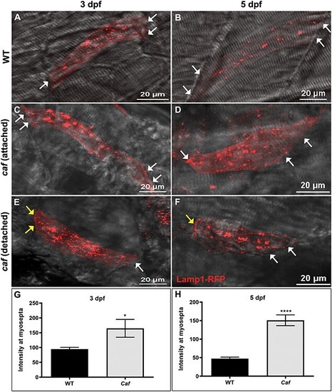
Lysosome distribution in caf mutants. (A–B) Lamp1-RFP expression in WT embryos shows that lysosomes are distributed throughout the cytoplasm in myofibers but not at myosepta (white arrows) at 3 and 5 dpf. (C–F) Lysosomes redistribute partially to the myosepta (white arrows) in caf mutants at 3 dpf (n = 10 fibers) and 5 dpf (n = 25 fibers), in both attached and detached fibers. Yellow arrows indicate where fibers have detached from the myosepta. (G–H) Lamp1-RFP fluorescence intensity at the myosepta was significantly increased in caf mutants at 3 dpf (*, P = 0.035) and 5 dpf (****, P < 0.0001). Bars represent mean ± SEM.
|