Figure 6
- ID
- ZDB-FIG-230313-7
- Publication
- Schaefer et al., 2023 - A low-cost smartphone fluorescence microscope for research, life science education, and STEM outreach
- Other Figures
- All Figure Page
- Back to All Figure Page
|
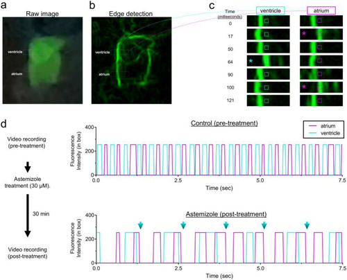
Glowscope detection of drug-induced cardiac arrythmia. (a–b) Still images from a glowscope video (acquired on an iPhone 11 Pro) show atrium and ventricle chambers in a 3 dpf Tg(myl7:EGFP) zebrafish embryo. Images show either the raw (unedited) view of heart fluorescence directly from smartphone video recording (a) or a processed view of the same heart after edge detection was performed on a computer using Fiji (b). Blue and magenta rectangles show the regions of ventricle and atrium chambers used for high-magnification viewing and analysis in (c–d). (c) Images show a timecourse (top to bottom) of ventricle (left) or atrium (right) chamber movements detected by the glowscope (corresponding to boxed regions in panel b). The small, dash-boxed regions of interest were used to monitor fluorescence intensity changes over time. Asterisks highlight frames where the heart chamber wall has moved into the region of interest with respect to the prior frame. (d) Graphs of fluorescence intensity vs. time plotted for atrium and ventricle chamber reveals oscillating chamber movements. Measurements within the same embryo (N = 1) were separately acquired and analyzed before (upper) and after astemizole treatment (lower). The maximum fluorescence intensity (in arbitrary units) within the dash-boxed region of interest shown in panel (c) was obtained and plotted for each time frame of the video recording, resulting in a fluorescence peak each time the chamber fluorescence entered and occupied the boxed region of interest. Note the differences in both heart rate between control and astemizole-treated hearts as well as the arrhythmic 2:1 atrial:ventricular beat pattern induced by astemizole treatment (lower plot, 30 µM). Blue arrows in panel (d) show missed ventricular beats. Videos available in Supplementary Video S1. |

