FIGURE

Figure 2

ID
ZDB-FIG-221214-230
Publication
Lee et al., 2022 - Evaluation of a Rapid and Simple Method for Assessing Retinal Vessel Structures in Adult Zebrafish
Other Figures
All Figure Page
Back to All Figure Page
Figure 2

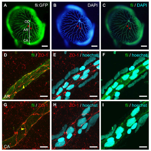
The cellular structure of whole-mount retinal vessels isolated from adult transgenic zebrafish, Tg[fli1:EGFP]. The vessel-specific expression of GFP (A) and nuclear staining with DAPI (B) in the retinal vasculature. (C) Merged image of (A,B). Retinal vessels from the optic disc (OD) branched out into the arterial region (AR) and then the capillary area (CA). Asterisks indicate membrane regions between blood vessels. (DI) Immunofluorescent labeling of tight junction protein (ZO-1, red), nuclear staining (Hoechst, blue), and GFP expression (green) in different retinal vessel regions; arterial region (DF) and capillary area (GI). Endothelial cells are elongated and their long axis is parallel to the line of the blood vessel. Arrows show cellular junctions between endothelial cells. Scale bars; 150 μm for (AC), 20 μm for (DF), and 10 μm for (GI).

Expression Data

Expression Detail
Antibody Labeling
Phenotype Data

Phenotype Detail
Acknowledgments
This image is the copyrighted work of the attributed author or publisher, and ZFIN has permission only to display this image to its users. Additional permissions should be obtained from the applicable author or publisher of the image. Full text @ Int. J. Mol. Sci.