FIGURE

Figure 2

ID
ZDB-FIG-221214-211
Publication
Lee et al., 2022 - Evaluation of Cisplatin-Induced Pathology in the Larval Zebrafish Lateral Line
Other Figures
All Figure Page
Back to All Figure Page
Figure 2

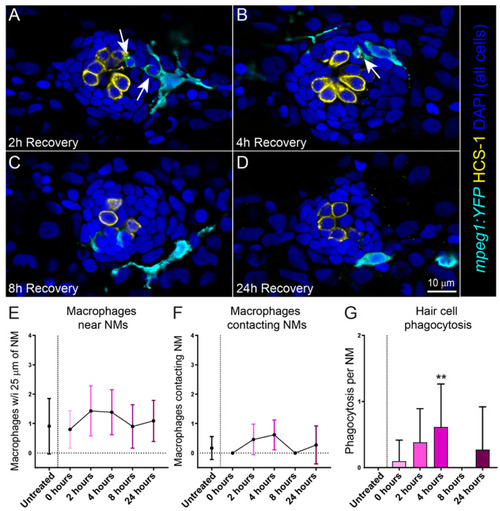
Macrophages respond to cisplatin-induced injury and phagocytose dying hair cells. (AD) Single z-section images taken from 15 µm-depth confocal stacks of neuromasts in fish following treatment with 250 µM cisplatin. Macrophages are labeled with YFP (cyan), hair cells with an antibody to Otoferlin (HCS-1; yellow) and all cell nuclei are labeled with DAPI (blue). Arrows indicate macrophage phagocytosis of dying hair cells. Note that the macrophages at 2- and 4-h recovery (A,B) have internalized otoferlin-labeled hair cell debris (yellow) as well as pyknotic nuclei (blue). At later time points (C,D) macrophages are in proximity of neuromasts but no longer phagocytosing hair cell debris (E,F). Quantification of macrophages in proximity to neuromasts (E) and contacting neuromasts (F) following cisplatin. There was no observed difference between cisplatin treated and control. (G) Quantification of phagocytotic events in cisplatin treated neuromasts, which peaked 4 h post cisplatin treatment (Dunn’s multiple comparisons test, ** adjusted p = 0.0636). Bars = SD. Data obtained from 10–14 fish/treatment group.

Expression Data

Expression Detail
Antibody Labeling
Phenotype Data
Fish:
Condition:
Observed In:
Stage: Day 6

Phenotype Detail
Acknowledgments
This image is the copyrighted work of the attributed author or publisher, and ZFIN has permission only to display this image to its users. Additional permissions should be obtained from the applicable author or publisher of the image. Full text @ Int. J. Mol. Sci.