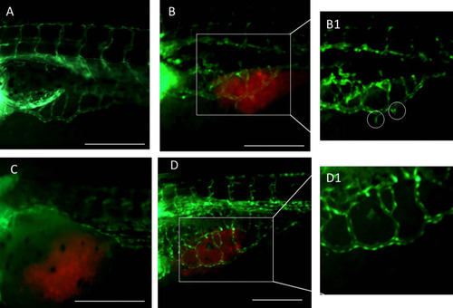
FIGURE

Fig. 6

ID
ZDB-FIG-220912-66
Publication
Moret et al., 2022 - Biodegradable nanoparticles combining cancer cell targeting and anti-angiogenic activity for synergistic chemotherapy in epithelial cancer
Other Figures
All Figure Page
Back to All Figure Page
Fig. 6

Vasculature analysis of fli1a:EGFP embryos after two days of treatment with nanoparticles (NPs) ([DTX] = 2.5 ng/animal). Fluorescence microscopy images of untreated embryos without (A) or with KB xenografted cells (B); embryos with KB xenografted cells and treated with free DTX (C) or DTX-DBLFol/aFLT1 (D). B1 is a magnification showing the presence of vascular branches (white circles) sprouting toward tumor masses in untreated embryos. In the magnification, D1 vascular branches are not visible due to the anti-angiogenic effect exerted by DTX-DBLFol/aFLT1. Scale bar: 250 μm. Red: tumor cells stained with DiI; green: zebrafish embryo vasculature

Expression Data

Expression Detail
Antibody Labeling
Phenotype Data

Phenotype Detail
Acknowledgments
This image is the copyrighted work of the attributed author or publisher, and ZFIN has permission only to display this image to its users. Additional permissions should be obtained from the applicable author or publisher of the image. Full text @ Drug Deliv Transl Res