
Fig. 6
- ID
- ZDB-FIG-220908-28
- Publication
- Ma et al., 2022 - Ercc2/Xpd deficiency results in failure of digestive organ growth in zebrafish with elevated nucleolar stress
- Other Figures
- All Figure Page
- Back to All Figure Page
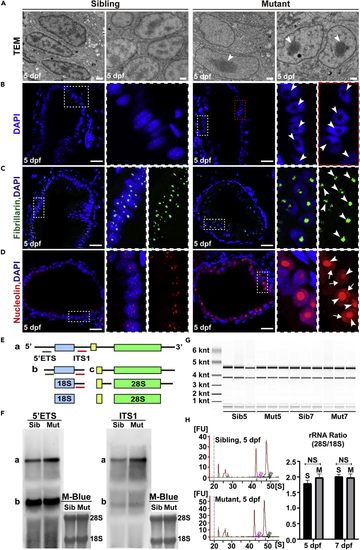
Perturbed rRNA synthesis results in nucleolar stress (A) Transmission electron microscopy images of intestinal endothelial cell (IEC) nuclei in ercc2/xpd mutants and siblings at 5 dpf. Arrowheads point to enlarged nucleoli. Scale bars, 1 μm. (B–D) DAPI staining and immunostaining of the nucleolar markers fibrillarin and nucleolin in IECs in ercc2/xpd mutants and siblings at 5 dpf. Areas of dashed boxes are magnified. Arrowheads point to enlarged nucleoli, arrows indicate the translocation of nucleolar proteins to the nucleoplasm. Scale bars, 20 μm. See also Figure S7. (E) Schematic diagram showing stepwise processing of the pre-rRNA transcript. a-c corresponds to the rRNA intermediates. 5′ETS and ITS1 probes are indicated with green and red lines, respectively. ETS, external transcribed spacer; ITS, internal transcribed spacer. (F) Northern blot using 5′ETS and ITS1 probes to detect precursor forms of rRNA in ercc2/xpd mutants and siblings at 5 dpf. Methylene blue staining was used as a loading control. (G and H) Representative E-Bioanalyzer analysis and measurement of the 28S/18S rRNA ratio in ercc2/xpd mutants (M) and siblings (S) at 5 and 7 dpf. Data are presented as mean ± SD, Student’s t test, NS, non-significant. |
| Antibodies: | |
|---|---|
| Fish: | |
| Anatomical Terms: | |
| Stage: | Day 5 |
| Fish: | |
|---|---|
| Observed In: | |
| Stage: | Day 5 |