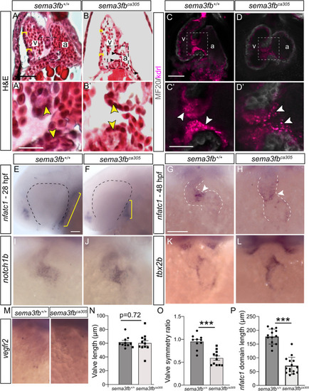
Disrupted heart valve development with the loss of Sema3fb. A, B Hematoxylin and eosin stained sections show the ventricle walls (yellow brackets) and valves (arrowheads within inset) of WT (A, A’) and sema3fbca305 embryos (B, B’) at 72 hpf. C, D Confocal projections of 72 hpf WT (C, C’) and mutant (D,D’) hearts immunolabeled by the myocardial marker MF20 (grey) on an endocardial-labeled transgenic line Tg(kdrl:mCherry). Brightness was increased in D’ to allow for comparison with C’. E, F nfatc1 ISH label in WT (E; N = 2, n = 13/13 normal) and sema3fbca305 (F, n = 13/15 short) 28 hpf embryos reveals a smaller endocardial domain (yellow bars). G, H nfatc1 concentration at the AVC (arrowheads) in WT (G, n = 21/21) and mutant (H, n = 16/17) 48 hpf fish. I-M AVC endocardial marker notch1b (I,J) (N = 2 WT n = 10/10; sema3fbca305 n = 10/10) and endocardial marker vegfr2 (M) (WT n = 10/12; sema3fbca305 n = 15/17) in 48 hpf hearts. Expansion of the tbx2b AVC cardiomyocyte marker in mutants (n = 7/8) that normally shows restricted expression at the AVC in WT hearts (n = 5/6) (K,L). N-P Quantitation of the: (1) length of the AV valve, as marked by mCherry in the Tg(kdrl:mCherry) background (N; N = 2, n = 10 sema3fb+/+, n = 12 sema3fbca305), (2) the ratio of the length of the valve present in the atrium to that measured in the ventricle at 72 hpf (O; p = 0.0003), and (3) the length of the nfatc1 domain (yellow bars, E and F) in 28 hpf WT (n = 13) and mutant (N = 15) embryos (P; p < 0.0001). Scale bars in A (A,B), A’ (A’,B’), C (C,D), C’ (C’,D’), E (E,F), G (G-M) are 50 µm
|
