
Fig. 2
- ID
- ZDB-FIG-220819-6
- Publication
- Hughes et al., 2022 - Clonal behaviour of myogenic precursor cells throughout the vertebrate lifespan
- Other Figures
- All Figure Page
- Back to All Figure Page
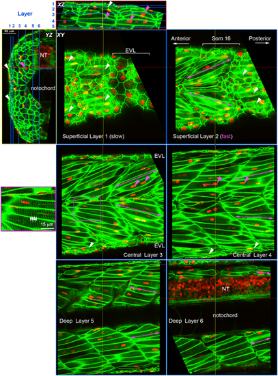
Cell content of the growing zebrafish somite. Larvae expressing membrane-localised EGFP and injected with RNA encoding H2B-mCherry were selected for brightness of both labels and scanned live at high resolution using a sub-Airy pinhole. Three orthogonal views are shown, with blue lines on XZ and YZ projections indicating the six sagittal XY planes. Red and yellow lines indicate the XZ and YZ planes, respectively. Note that EGFP labels the transverse T-tubule system (white arrows) in muscle fibres at regular 2 µm spacing orthogonal to the long axis of the fibre (purple box, magnified at left). Mononucleated fibres parallel to the anteroposterior axis on the surface of the myotome (white arrowheads) are superficial slow fibres. Fast fibres with multiple nuclei (magenta arrowheads) show different orientation depending on the depth within the myotome (magenta arrows); superficially they ‘point’ anteriorly in the sense that fibres in epaxial and hypaxial myotome project so that their anterior ends are closer to the horizontal myoseptum than their posterior ends (Superficial Layer 2 and Central Layer 3). In the medial myotome, by contrast, fibres have the converse orientation, so that epaxial and hypaxial fibres ‘point’ posteriorly (Deep Layer 5 and 6). NT, neural tube. |