
FIGURE
Fig. 1
- ID
- ZDB-FIG-220603-52
- Publication
- Xu et al., 2022 - Orphan G-Protein Coupled Receptor GPRC5B Is Critical for Lymphatic Development
- Other Figures
- All Figure Page
- Back to All Figure Page
Fig. 1
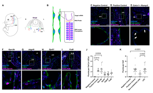
Figure 1. In situ localization of lymphatic endothelial cell-enriched GPCR expression. (A) Schematic diagram of E14.5 transverse section of mouse embryo depicting the sterotypic positioning of carotid artery (CA), jugular vein (JV) and jugular lymph sac (JLS). (B) Schematic diagram of RNAscope probe design, illustrating how a single mRNA molecule is amplified into a visible fluorophore puncta within a cell. (C–I) RNAscope images for indicated probes (magenta), Hoechst nulcei (blue) and the lymphatic maker LYVE-1 or Calcrl in panel (E) (green). Probe names are listed at top of images. Lower images are higher magnification views of the white dashed inset. White arrows in (E) point to lymphatic endothelial cells with double-labeling of the lymphatic GPCR, Calcrl (green) and Ramp3 (magenta). Scale bars = 50 µm. (J) RNAscope quantification for each orphan receptor probe in lymphatic endothelial cells and (K) RNAscope quantification for Gprc5b in various tissue types. Puncta per cell is a relative measure of the number of mRNA transcripts for the target gene. Non-endothelial cells (non-ECs) represent all background cells. LEC = lymphatic endothelial cells. Significance calculated using Kruskal–Wallis nonparametric analysis of variance with Dunn’s multiple comparisons test. p-values equal to or less than 0.05 are shown.
|
Expression Data
Expression Detail
Antibody Labeling
Phenotype Data
Phenotype Detail
Acknowledgments
This image is the copyrighted work of the attributed author or publisher, and
ZFIN has permission only to display this image to its users.
Additional permissions should be obtained from the applicable author or publisher of the image.
Full text @ Int. J. Mol. Sci.