
FIGURE
Fig. 5
- ID
- ZDB-FIG-220527-5
- Publication
- Gao et al., 2022 - Accumulation of Lipid Droplets in a Novel Bietti Crystalline Dystrophy Zebrafish Model With Impaired PPARα Pathway
- Other Figures
- All Figure Page
- Back to All Figure Page
Fig. 5
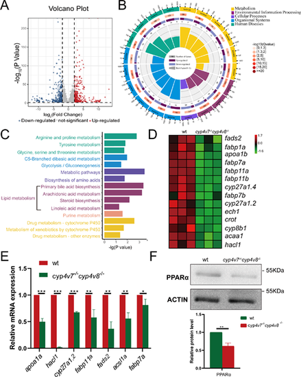
Downregulation of the PPARα pathway was involved in cyp4v7/cyp4v8 DKO zebrafish RPE cells. (A) Volcano plot of differentially expressed genes in cyp4v7−/−cyp4v8−/− zebrafish compared with WT. (B) KEGG circle enrichment analysis of the differentially expressed genes. The first lap indicates the top 20 KEGG terms. The second lap represents the gene numbers in the genome backdrop. The third circle represents the ratio of the upregulated genes (dark purple) and downregulated genes (light purple). The fourth circle represents the enrichment element of each KEGG term. (C) KEGG enriched metabolism pathways (P < 0.05) are shown. (D) Expression patterns of genes in PPARα signaling pathway in WT and cyp4v7−/−cyp4v8−/− zebrafish RPE cells are shown in the heatmap. (E) Validation of the expression of genes in PPARα pathway by qPCR in cyp4v7 and cyp4v8 DKO zebrafish RPE cells at seven months after fertilization. (F) Detection of the protein level of PPARα in WT and cyp4v7−/−cyp4v8−/− zebrafish RPE cells by Western blot. The results are shown as mean ± SD. ***P < 0.001; **P < 0.01; *P < 0.05.
|
Expression Data
| Genes: | |
|---|---|
| Antibody: | |
| Fish: | |
| Anatomical Term: | |
| Stage: | Adult |
Expression Detail
Antibody Labeling
Phenotype Data
| Fish: | |
|---|---|
| Observed In: | |
| Stage: | Adult |
Phenotype Detail
Acknowledgments
This image is the copyrighted work of the attributed author or publisher, and
ZFIN has permission only to display this image to its users.
Additional permissions should be obtained from the applicable author or publisher of the image.
Full text @ Invest. Ophthalmol. Vis. Sci.