FIGURE

Figure 3

ID
ZDB-FIG-220520-4
Publication
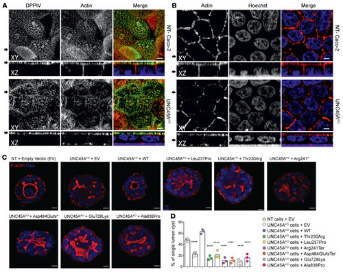
Duclaux-Loras et al., 2022 - UNC45A deficiency causes microvillus inclusion disease-like phenotype by impairing myosin VB-dependent apical trafficking
Other Figures
All Figure Page
Back to All Figure Page
Figure 3

Disrupted enterocyte architecture in <italic toggle='yes'>UNC45A</italic> deficiency.

(A) Confocal images of polarized NT control and Unc45AKO Caco-2 cells grown on a filter and stained for an apical brush border marker DPPIV and actin. (B) Actin staining in polarized Caco-2 cells. Nuclei were visualized with HOECHST. Arrows on the left mark the corresponding XY and XZ planes. Scale bars: 20 μm. Panels A and B were from the same experiment. (C) NT control and UNC45AKO Caco-2 cells complemented or not with WT or mutant alleles were cultured in 3D for 5 days to form cysts. Nuclei are stained with Nucblue (blue); actin is stained with phalloidin AF 455 (red). Single confocal sections through the middle of the cyst are shown. Scale bars: 10 μm. (D) Single-lumen cysts were counted in each experiment. Results from 3 independent experiments (35 cysts each) are shown, 1-way ANOVA. ****P < 0.0001.

Expression Data

Expression Detail
Antibody Labeling
Phenotype Data

Phenotype Detail
Acknowledgments
This image is the copyrighted work of the attributed author or publisher, and ZFIN has permission only to display this image to its users. Additional permissions should be obtained from the applicable author or publisher of the image. Full text @ Journal of Clin. Invest.