
Figure 3—figure supplement 1.
- ID
- ZDB-FIG-220324-37
- Publication
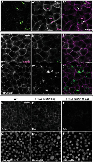
- Saraswathy et al., 2022 - The E3 Ubiquitin Ligase Mindbomb1 controls planar cell polarity-dependent convergent extension movements during zebrafish gastrulation
- Other Figures
-
- Figure 1
- Figure 1—figure supplement 1.
- Figure 1—figure supplement 2.
- Figure 1—figure supplement 3.
- Figure 2
- Figure 3
- Figure 3—figure supplement 1.
- Figure 3—figure supplement 2.
- Figure 3—figure supplement 3.
- Figure 3—figure supplement 4.
- Figure 3—figure supplement 5.
- Figure 4
- Figure 4—figure supplement 1.
- Figure 5
- Figure 5—figure supplement 1.
- All Figure Page
- Back to All Figure Page
( |