
Fig. 7
- ID
- ZDB-FIG-220301-13
- Publication
- Donati et al., 2021 - Planar polarization of cilia in the zebrafish floor-plate involves Par3-mediated posterior localization of highly motile basal bodies
- Other Figures
- All Figure Page
- Back to All Figure Page
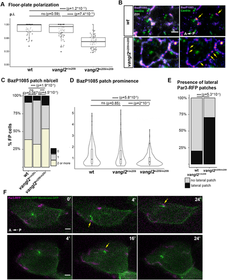
Par3 clustering and localization in vangl2m209 FP. (A) p.i. of vangl2m209/m209 determined from immunostaining data. Wild type: two embryos, 49 cells; vangl2m209/+: three embryos, 66 cells; vangl2m209/m209: five embryos, 57 cells. Box boundaries represent the first and third quartiles of the distribution, and boxplot whiskers span 1.5 times the interquartile range of the distribution; each dot represents one cell. ns P>0.05; ****P<0.0001 (Wilcoxon rank sum test). (B) Immunostaining of phosphorylated Par3 (BazP1085 antibody) in vangl2+/+ (wild type) and vangl2m209/m209 embryo FP at 18s. ZO1 staining was removed in the right images to reveal Par3 patches (yellow arrows). (C) Quantification of Par3 patch number per cell on ant/post membranes from immunostaining data as shown in B. ns P>0.05; ***P<0.001 (Fisher's exact test). (D) Prominence of Par3 patches (BazP1085 antibody) in wt and vangl2m209/m209 mutant embryo FP at 18s. ns P>0.05; ***P<0.001; ****P<0.0001 (Wilcoxon rank sum test). In A-D, vangl2+/+: seven embryos, 186 cells; vangl2m209/+: five embryos, 112 cells; vangl2m209/m209: seven embryos, 129 cells. (E) Percentage of cells displaying a lateral Par3-RFP patch in live imaging (such as in F). vangl2+/+ and vangl2m209/+: 16 embryos, 45 cells; vangl2m209/m209: seven embryos, 17 cells. ***P<0.001 (Fisher's exact test). (F) Images from two movies of 5s vangl2m209/m209 embryos mosaically injected with Par3-RFP, Centrin-GFP and Membrane-GFP mRNA at the 16- to 32-cell stage. Yellow arrows indicate contact events between lateral Par3 patches and BBs. Scale bars: 2 µm. |