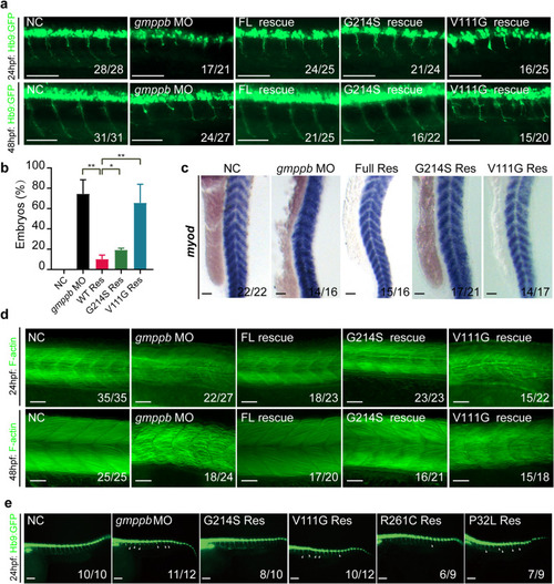
GMPPB activity is required for muscle and neuronal development (a-b) GMPPB V111G mutant with decreased activity fails to rescue axonal phenotype in gmppb MO-injected zebrafish. Morphology of CaP axons from embryos at 24 and 48 hpf that were injected with control MO (NC), or gmppb MO alone or together with mRNA encoding GMPPB WT or its mutants at one-cell stage of the Tg [hb9: GFP]ml2 transgenic zebrafish embryos. Scale bar: 100 μm (a). Statistical results of the embryos with abnormally branched axons (b). c-d GMPPB V111G mutant fails to rescue muscle defects caused by Gmppb KD. Expression of myod in zebra fish injected with control MO (NC), or gmppb MO alone or together with mRNA encoding GMPPB WT or its mutants. Scale bar: 100 μm (c). Phalloidin staining of filamentous actin (green) in zebra fish injected with control MO (NC), or gmppb MO alone or together with mRNA encoding GMPPB WT or its mutants at one-cell stage. Scale bar: 100 μm (d). e The enzymatic activity of GMPPB mutant positively correlates its ability in rescuing axonal phenotype. Morphology of CaP axons from embryos at 24 and 48 hpf that were injected with control MO (NC), or gmppb MO alone or together with different mRNA at one-cell stage of the Tg [hb9: GFP]ml2 transgenic zebrafish embryos. Scale bar: 80 μm. Mean ± SD, ****p < 0.0001; ***p < 0.001; **p < 0.01; *p < 0.05. p values were calculated using one-way ANOVA, Tukey’s multiple comparisons test
|
