
Figure 2.
- ID
- ZDB-FIG-211207-91
- Publication
- Kenney et al., 2021 - A 3D adult zebrafish brain atlas (AZBA) for the digital age
- Other Figures
- All Figure Page
- Back to All Figure Page
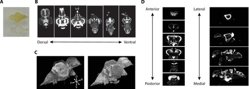
(A) Image of adult zebrafish brain samples before (top) and after (bottom) clearing using iDISCO+. (B) Example TO-PRO-stained images from a single sample acquired in the horizontal plane during light-sheet imaging. (C) Three-dimensional volumes generated from a set of light-sheet images from an individual brain visualized using a maximum intensity projection (left) and exterior volume (right). (D) Coronal (left) and sagittal (right) views of an individual brain generated from a single three-dimensional volume.
|