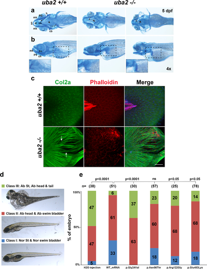
Fig. 4. Cranial cartilage patterns observed in uba2-/- zebrafish and rescue of uba2 mutant phenotype with human UBA2 messenger RNA (mRNA). (a, b) Brightfield ventral and lateral views of cartilage stained uba2 in wild type (WT) and homozygous mutant fish are shown in top and bottom panels, respectively. Closeups of pectoral fin cartilage phenotype are shown in inserts in the bottom panel highlighted by black dashed boxes on lateral views. an anterior, cb ceratobranchials 1–4, ch ceratohyal, ep ethmoid plate, h hypohyal, hs hyosymplectic, mk Meckel’s cartilage, pq palatoquadrate. (c) Z-stack images of uba2 zebrafish median fins stained with Col2a (green), Rhodamine-Phalloidin (red), and Dapi (blue). Arrows are used to show the gaps between actinotrichia fibers. Scale bar: 50 µm. (d) Suppression of uba2 in zebrafish produces an abnormal phenotype that is classified into three categories. (e) Proportions of uba2-/- zebrafish embryos representing each phenotype category after injecting with WT or mutation harboring human UBA2 mRNA. Landmark abbreviations: Ab abnormal, Nor St normal structure, ns not significant. Chi Square test p values are shown above the phenotypes for each rescue experiment.
|
