
Figure 4
- ID
- ZDB-FIG-210902-229
- Publication
- Hiyoshi et al., 2021 - Two-Photon Laser Ablation and In Vivo Wide-Field Imaging of Inferior Olive Neurons Revealed the Recovery of Olivocerebellar Circuits in Zebrafish
- Other Figures
- All Figure Page
- Back to All Figure Page
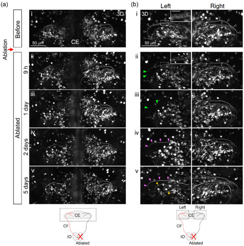
Structural changes of climbing fibers (CFs) in the cerebellar region induced by the two-photon laser ablation of the inferior olive. (a,b) Three-dimensional dorsal view of climbing fibers. The area of observations is shown by the dotted line in the schematic diagram. Before ablation: i, after ablation: ii (9 h), iii (1 day), iv (2 days), and v (5 days). (b) A similar region as (a), but the orientation is slightly different: A high-magnification image is inserted in the upper right corner in (b-i) (before) and (b-ii) (after 9 h). Scale bar: 10 μm. Black arrowheads in (b-i) indicate the bundle of CFs. Green arrowheads in (b-ii,iii) indicate the defected CFs after laser ablation. Red and yellow arrowheads (b-iv,v) indicate newly emerged CFs. |