|
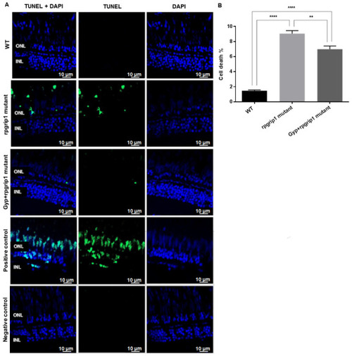
Significant decreases were found in cone apoptosis in rpgrip1 mutant zebrafish treated with Gyp. (A) Retinal sections of wildtype (WT), untreated (UT) rpgrip1 mutant and Gyp-treated rpgrip1 mutant zebrafish at 6 mpf were stained with TUNEL reagents. Nuclei of apoptotic cone cells were stained in green. DAPI labelled nuclei were stained in blue. (B) Quantification of apoptotic cells in above retinal sections were compared between the wildtype, rpgrip1 mutant and Gyp-treated rpgrip1 mutant zebrafish. Statistical comparisons between individual groups were carried out using one-way ANOVA followed by Bonferroni’s test. Data are displayed as mean ± SEM (n = 6 animals of each group). ** p < 0.01, **** p < 0.0001. INL, inner nuclear layer; ONL, outer nuclear layer.
|