FIGURE

Fig 3

ID
ZDB-FIG-210714-23
Publication
Pottie et al., 2021 - Loss of zebrafish atp6v1e1b, encoding a subunit of vacuolar ATPase, recapitulates human ARCL type 2C syndrome and identifies multiple pathobiological signatures
Other Figures
All Figure Page
Back to All Figure Page
Fig 3

Effect of loss of <italic toggle='yes'>atp6v1e1b</italic> on the non-canonical function of v-ATPases.

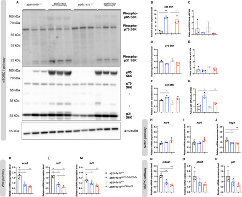
(A) Phosphorylated ribosomal protein S6 kinase 1 (S6K1) (upper panel) and total S6K1 (lower panel) were detected in protein lysates of whole atp6v1e1b-deficient zebrafish and their corresponding WT controls at 4 dpf by immunoblotting. Asterisks indicate non-related, cross-reactive bands. Representative immunoblots are shown. We confirmed equal loading by staining for α-tubulin. (B-D-F) Band intensities of chemiluminescent signals of non-phosphorylated proteins were quantified with ImageJ and normalized to WT controls. (C-E-G) Band intensities of chemiluminescent signals of phosphorylated proteins were quantified with ImageJ. The ratio of phospho-p85-S6K to total p85-S6K, phospho-p70-S6K to total p70-S6K, and phospho-p31-S6K to total p31-S6K was normalized to WT controls. Data are expressed as mean ± SD from 3 biological replicates in B-G. (H-P) Gene expression of Notch-target genes (her4, her6, and hey2), Wnt-target genes (axin2, tcf7, and lef1), and AMPK-target genes (prkaa1, ptch1, and gli1) were investigated by RT-qPCR. Data are expressed as mean ± SD from 5 biological replicates in H-P.

Expression Data

Expression Detail
Antibody Labeling
Phenotype Data

Phenotype Detail
Acknowledgments
This image is the copyrighted work of the attributed author or publisher, and ZFIN has permission only to display this image to its users. Additional permissions should be obtained from the applicable author or publisher of the image. Full text @ PLoS Genet.