FIGURE

FIGURE 6

ID
ZDB-FIG-210710-25
Publication
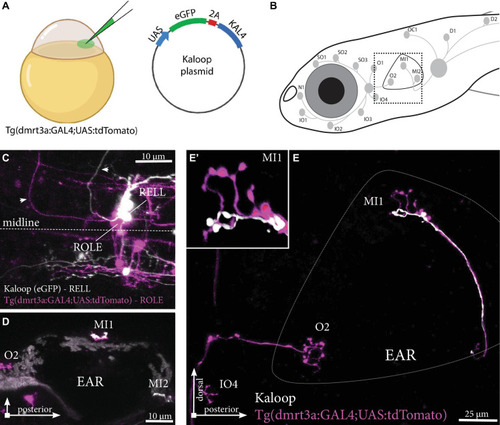
Manuel et al., 2021 - Characterization of Individual Projections Reveal That Neuromasts of the Zebrafish Lateral Line are Innervated by Multiple Inhibitory Efferent Cells
Other Figures
All Figure Page
Back to All Figure Page
FIGURE 6

Tg(dmrt3a:GAL4;UAS:tdTomato) injected with Kaloop plasmid reveal multiple inhibitory efferent cells innervating a single neuromast. (A) One-cell stage embryos of Tg(dmrt3a:GAL4;UAS:tdTomato) were injected with Kaloop plasmid to generate larvae with sparsely labeled tdTomato and eGFP inhibitory efferents. (B) Schematic overview of the area shown in (E). (C–D) Confocal images showing the view of sparsely labeled inhibitory efferent cells in their nuclei. A ROLE (tdTomato-positive; magenta) and a RELL (eGFP-positive; white) cell were identified (C) and their projections traced across the lateral line (D) dorsal view and (E) lateral view of the inhibitory efferent projections at the level of the ear. In this case, both efferent cells were connected to the same neuromast located on the ear (MI1) [(E’) depicts the MI1 neuromast at higher magnification]. Age of larvae: 5 dpf. ROLE, the rhombencephalic octavolateral efferent neuron; RELL, the rhombencephalic efferent neuron to the lateral line. (A) was partially created with BioRender.com

Expression Data

Expression Detail
Antibody Labeling
Phenotype Data

Phenotype Detail
Acknowledgments
This image is the copyrighted work of the attributed author or publisher, and ZFIN has permission only to display this image to its users. Additional permissions should be obtained from the applicable author or publisher of the image. Full text @ Front. Neuroanat.