
Figure 2
- ID
- ZDB-FIG-210628-55
- Publication
- Lee et al., 2021 - Zebrafish, an In Vivo Platform to Screen Drugs and Proteins for Biomedical Use
- Other Figures
- All Figure Page
- Back to All Figure Page
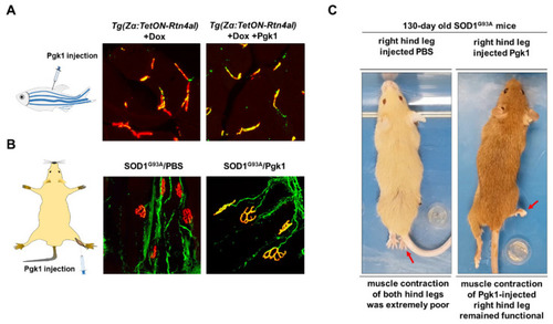
Intramuscular injection of Pgk1 could reduce the NMJ denervation occurring in the ALS animal models of zebrafish and mouse. (A) ALS-like adult zebrafish model. Left: diagram illustrates the muscle injection of Pgk1 into adult zebrafish; middle: after Doxycycline (Dox) was treated for one week, a few number of axonal motor neurons (MNs; labeled with green fluorescence signal) co-localized with motor end plate (labeled with red fluorescent signal) presented in yellow fluorescent signal was observed in the ALS-like zebrafish strain Tg(Zα:TetON-Rtn4al), indicating that NMJ denervation, a pathogenesis indicator at early stage of ALS, seriously occurred in the Rtn4al/NogoA-overexpressed zebrafish; right: Pgk1 was injected into the muscle of Dox-treated adult zebrafish, and the number of axonalMNs (labeled with green fluorescence signal) co-localized with motor end plate (labeled with red fluorescent signal) presented in yellow fluorescent signal was increased, indicating extracellular injection of Pgk1 could reduce the denervation and maintain the complete NMJ structure in the Rtn4al/NogoA-overexpressed zebrafish. (B) ALS (SOD1G93A) mice model. Left: diagram illustrates that the solution was injected into the gastrocnemius of right hind leg of ALS mice; middle: phosphate buffer saline (PBS) served as control was injected into the gastrocnemius of right hind leg of ALS mice, and the denervation of axonal MNs was examined. A few numbers of axonal MNs (labeled with green fluorescence signal) co-localized with motor end plate (labeled with red fluorescent signal) presented in yellow fluorescent signal was observed in the 75-day-old ALS mice; right: Pgk1 was injected into the gastrocnemius of right hind leg of ALS mice at 60 days old, and the denervation of axonal MNs was examined at 75 days old. The number of NMJ with complete structure presented a yellow fluorescent signal was increased compared to that of control group. (C) The first shot of Pgk1 was injected into the gastrocnemius of right hind leg of SOD1G93A ALS-mice at 60 days old, followed by the other shot for every 15 days until 120s days old. The treated ALS-mice were observed at 130 days old. Left: both hind legs of ALS-mice injected with PBS (control) were completely paralyzed. Right: the right hind leg injected with Pgk1 could keep contraction and movable, while the left hind leg was paralyzed. Figure modified from [106]. |